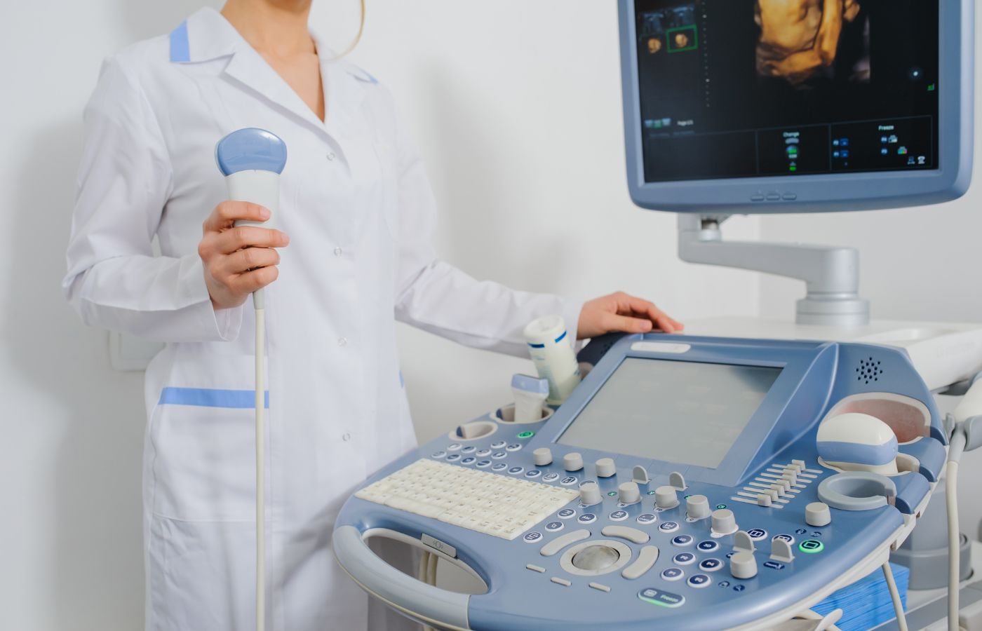
When it comes to diagnosing conditions related to the chest and pain, imaging plays a crucial role in determining the root cause. Among the many diagnostic options, USG chest, or chest ultrasound, is emerging as a powerful tool alongside conventional imaging methods like X-ray and CT scan. This blog explores the purpose of chest ultrasound, how it compares to an X-ray, and where to find the best diagnostic centre near you .
USG (Ultrasonography) of the chest is a non-invasive imaging technique that uses high-frequency sound waves to create ultrasound images of structures inside the chest. Unlike X-rays that use ionizing radiation, USG is safe, radiation-free, and suitable for repeated use—especially in sensitive individuals such as children, pregnant women, and critically ill patients.
A chest ultrasound serves various purposes, particularly in assessing soft tissue structures. It is frequently used in:
In many cases, chest ultrasound is preferred over X-rays due to its superior ability to detect small fluid collections and soft tissue abnormalities.
Each imaging modality has its strengths. Here is a quick comparison:
Criteria USG Chest X-ray Radiation No Yes Best For Fluid, soft tissues Bone, lung fields Accuracy in Pleural Effusion High Moderate Bedside Availability Yes Limited Cost Affordable Affordable Limitations Cannot penetrate air-filled structures Poor soft tissue contrastWhile X-rays are great for getting a general view of the lungs and bones, ultrasound imaging offers better clarity in identifying fluid collections and guiding interventional procedures. In emergency and ICU settings, USG is invaluable due to its bedside availability.
If you're experiencing any of these symptoms, searching for an ultrasound near me or ultrasound scanning near me is the first step toward accurate diagnosis.
A doctor may recommend a USG chest in the following scenarios:
Because of its real-time imaging capability, USG is especially useful in intensive care units, emergency departments, and outpatient diagnostic settings.
Advantages of USG ChestDespite its limitations, when performed by skilled radiologists, chest ultrasound offers precise, targeted diagnostics.
How to Prepare for a Chest Ultrasound Minimal preparation is required:The scan typically takes 10-20 minutes and is completely non-invasive.
Chest ultrasound (USG chest) is a safe, accurate, and efficient imaging option for diagnosing a wide range of chest-related conditions. Whether you’re experiencing unexplained chest and pain chest and pain, suspect a lung infection, or need monitoring for an ongoing condition, a chest ultrasound can provide the clarity you need.
Looking for quality diagnostic services? Visit Diagnopein Diagnostic Centres, one of the best diagnostic centres near you, offering advanced ultrasound scanning near you with expert care and accurate reports.
Take charge of your health today—book your USG chest scan and breathe easy knowing you're in expert hands.