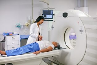
The Role of MRI in Parkinson’s Disease Diagnosis
MRI Brain Parkinson Protocol helps in the differential diagnosis of Parkinson’s disease. While the disease is primarily diagnosed based on clinical symptoms (tremors, rigidity, bradykinesia), imaging techniques like MRI are invaluable in excluding other conditions that might mimic Parkinson’s, such as:
1] Multiple System Atrophy (MSA): An MRI can help distinguish MSA from Parkinson’s by showing certain atrophy patterns in the brainstem and cerebellum, which are not typically seen in Parkinson’s.
2] Progressive Supranuclear Palsy (PSP): The MRI can identify characteristic changes in the midbrain that are often seen in PSP but are absent in Parkinson’s disease.
3] Normal Pressure Hydrocephalus (NPH): MRI can reveal the enlargement of the ventricles in the brain, which is typical of NPH and helps differentiate it from Parkinson’s disease.
Furthermore, while MRI is not the definitive diagnostic tool for Parkinson’s disease, it can support the clinical assessment and help clinicians evaluate the extent of brain degeneration, aiding in the management and monitoring of the disease.
Monitoring Disease Progression
MRI Brain Parkinson Protocol is also helpful in tracking the progression of Parkinson's disease over time. As Parkinson’s advances, changes in the brain structures, particularly in the substantia nigra and basal ganglia, become more pronounced. MRI scans can track these changes and offer valuable information on how the disease is evolving. This can be critical in adjusting treatment plans and improving patient outcomes.
Benefits of MRI in Parkinson’s Diagnosis
1] Non-invasive: Unlike biopsy or other invasive procedures, MRI is a non-invasive imaging technique that poses minimal risk to patients.
2] High Sensitivity: Specialized MRI techniques, like SWI and DTI, have shown high sensitivity in detecting early and subtle changes in the brain, helping in early diagnosis.
3] Disease Monitoring: MRI scans allow for the long-term tracking of disease progression, which can guide treatment decisions and interventions.
4] Exclusion of Other Conditions: MRI Brain Parkinson Protocol helps exclude other potential causes of movement disorders, allowing for a more accurate diagnosis.