


Diagnopein MRI Venography (Walkin DHPL) - CIMS Bhopal Centre in Bhopal
Diagnopein MRI Venography (Walkin DHPL) - CIMS Bhopal Centre in Bhopal
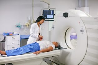
MRI Venography, also known as MR Venography (MRV), is a non-invasive imaging technique used to visualize the veins in the body, particularly those in the brain, legs, and abdomen. It employs the power of magnetic resonance imaging (MRI) to provide detailed pictures of venous structures, offering critical information for diagnosing various vascular conditions. Unlike traditional venography, which involves the injection of a contrast dye into the veins, MRI venography typically uses a magnetic field and radio waves to generate high-resolution images, and in some cases, contrast agents may be used to enhance the images.


