Purpose of 3D Sonography
1. Pregnancy and Fetal Imaging: One of the most common uses of 3D sonography is in obstetrics to capture detailed images of the developing fetus. It helps assess fetal development, detect abnormalities, and provide parents with a clearer view of the baby during pregnancy. 3D ultrasound allows for more detailed visualizations of the baby’s facial features, limbs, and overall growth, which can be difficult to capture with 2D ultrasound.
2. Diagnosis of Abnormalities: 3D ultrasound is often used to evaluate anatomical abnormalities in the fetus, including cleft lip, cleft palate, spina bifida, and heart defects. It can offer more detailed images of certain organs and structures that may be difficult to evaluate with traditional 2D scans.
3. Musculoskeletal Imaging: 3D sonography is also used in the assessment of musculoskeletal conditions. It can provide detailed images of muscles, tendons, and joints, helping diagnose injuries, tears, or degenerative changes that may not be clearly visible on 2D scans.
4. Cardiological Imaging: In cardiology, 3D ultrasound is used to capture a detailed view of the heart and its chambers, enabling doctors to assess heart function, detect abnormalities, and plan surgeries or treatments.
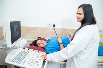
At Diagnopein, the best sonography centre in Mumbai, we offer cutting-edge 3D sonography imaging services. This advanced technology provides highly detailed, three-dimensional images of internal organs and structures, allowing for better diagnosis and treatment planning. Whether it's for pregnancy monitoring, examining organs, or assessing health conditions, our 3D sonography delivers clearer, more accurate results. Trust our expert team in Mumbai to offer the highest level of care with state-of-the-art equipment. Visit Diagnopein for precise and reliable 3D sonography images, enhancing your healthcare experience.
Advantages of 3D Sonography
1. Enhanced Clarity: Provides more detailed and realistic images of organs and structures, offering a clearer understanding of conditions.
2. Better Visualization of Fetal Anatomy: This can help detect physical abnormalities that might be missed on traditional 2D ultrasounds.
3. Non-invasive: Like traditional ultrasound, 3D sonography is a safe, non-invasive imaging method with no radiation exposure.
4. Real-time Imaging: Allows healthcare providers to view the structures in three dimensions during the procedure, offering better diagnostic capabilities.