Why is CT Abdomen & Pelvis with Contrast Important?
The addition of contrast dye in a CT scan significantly improves the quality of the images, making it easier for radiologists to identify abnormalities. Here are some reasons why this test is crucial:
1. Enhanced Detection of Tumors and Masses: The contrast dye helps distinguish between normal and abnormal tissues. It makes tumors, cysts, and other growths more visible, allowing for early detection and accurate assessment of size, shape, and location.
2. Evaluation of Inflammatory Conditions: For conditions like appendicitis, diverticulitis, and inflammatory bowel disease (IBD), the enhanced visibility from contrast dye helps in assessing the extent of inflammation and infection. This aids in diagnosing the cause of abdominal pain and planning appropriate treatment.
3. Identification of Vascular Abnormalities: The contrast dye outlines blood vessels, helping detect issues such as aneurysms, blood clots, and blockages. This is particularly important for diagnosing conditions like mesenteric ischemia, where blood flow to the intestines is compromised.
4. Assessment of Organ Function and Damage: The scan provides detailed images of the liver, kidneys, pancreas, spleen, and bladder, helping in the evaluation of conditions such as liver cirrhosis, pancreatitis, kidney stones, and bladder tumors. The use of contrast enhances the differentiation between normal and diseased tissue.
5. Guidance for Surgical Planning and Biopsies: In cases where surgical intervention is needed, the detailed images from a contrast-enhanced CT scan help surgeons plan procedures effectively. It is also used to guide needle biopsies, allowing precise targeting of abnormal tissue.
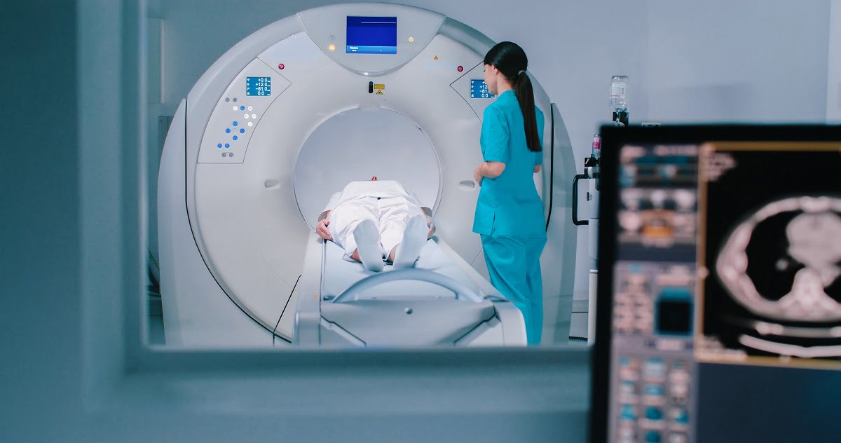
How is the CT Abdomen & Pelvis with Contrast Performed?
The procedure for a CT scan of the Abdomen and Pelvis with Contrast involves several steps:
1. Preparation:
Fasting: Patients are usually asked to fast for at least 4 to 6 hours before the scan to ensure clear images. Hydration: It is recommended to stay well-hydrated before the test to help flush the contrast dye out of the system afterward. Allergy Check: Patients should inform their healthcare provider of any known allergies, especially to iodine or shellfish, as these may indicate sensitivity to the contrast dye.
2. Administration of Contrast Dye:
The contrast dye is typically injected into a vein in the arm. In some cases, an oral contrast may also be given to help highlight the digestive tract. The patient may feel a warm sensation or a metallic taste in the mouth when the dye is administered, but this usually subsides quickly.
3. Positioning:
The patient lies on a motorized table, usually on their back. The table then moves slowly into the CT scanner, a large, donut-shaped machine. The patient may be asked to hold their breath for short periods to ensure clear images.
4. Imaging Process:
The scanner takes a series of X-ray images from different angles, which are processed by a computer to create detailed cross-sectional images of the abdomen and pelvis. The entire scan typically takes about 10 to 30 minutes.
5. Post-Scan Care:
After the scan, patients are advised to drink plenty of fluids to help flush the contrast dye from their system. Most people can resume their normal activities immediately after the scan.
Who Should Consider a CT Abdomen & Pelvis with Contrast?
A CT scan with contrast is recommended for patients experiencing symptoms or conditions where enhanced imaging can provide a clearer diagnosis:
1. Unexplained Abdominal Pain: For patients with severe or persistent abdominal pain, the use of contrast helps identify the underlying cause, such as infections, inflammation, or tumors.
2. Suspected Cancers or Masses: This scan is often used when there is a suspicion of cancer in the abdominal organs or pelvis, as it provides detailed information about the presence, size, and spread of tumors.
3. Evaluation of Trauma: In emergency situations involving abdominal injuries, such as from car accidents or falls, a contrast-enhanced CT scan helps identify internal bleeding, organ damage, and fractures.
4. Monitoring of Chronic Conditions: For patients with known conditions like liver disease, kidney problems, or inflammatory bowel disease, this scan helps monitor disease progression and evaluate treatment effectiveness.