Why is CT Brain with Contrast Important?
A CT brain with contrast is crucial for diagnosing various neurological conditions. The use of contrast allows for enhanced imaging, making it easier to identify problems that might not be visible on a non-contrast CT scan. Here are the main reasons why this test is important:
1. Detection of Brain Tumors: The contrast dye helps in distinguishing between normal tissue and abnormal growths. Tumors, cysts, and other masses in the brain appear more clearly, allowing healthcare providers to assess their size, location, and potential impact on surrounding structures.
2. Evaluation of Stroke: In cases of stroke, a CT scan with contrast can show areas of the brain that have been affected by a blockage or bleeding. The contrast helps visualize damaged brain tissue, blood vessels, and signs of ischemia (lack of blood flow), which is essential for determining treatment options.
3. Diagnosis of Infections: Conditions like brain abscesses, meningitis, or encephalitis can be evaluated using CT with contrast. The contrast highlights areas of infection or inflammation in the brain, aiding in diagnosis and helping to guide treatment decisions.
4. Assessment of Blood Clots and Hemorrhage: If there is a concern about internal bleeding or blood clots in the brain (such as from trauma or a hemorrhagic stroke), the contrast enhances the ability to detect these issues. It also helps in identifying the location and size of hemorrhages.
5. Chronic Neurological Conditions: For conditions such as multiple sclerosis or vascular malformations, a CT scan with contrast can provide valuable insight into the extent of disease progression and the effectiveness of treatment.
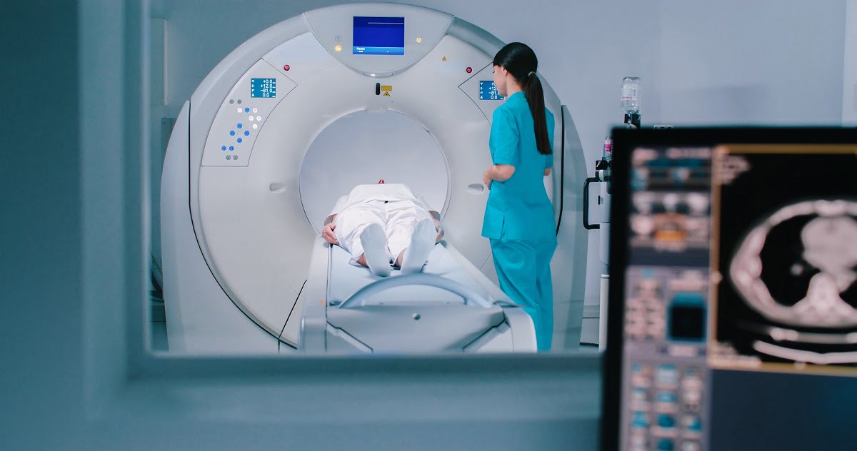
How is a CT Brain with Contrast Performed?
A CT brain with contrast follows a similar procedure to a regular CT scan, with the addition of contrast dye:
1. Preparation: Generally, no special preparation is required for a CT scan of the brain. However, patients may be asked to remove any metal objects (such as jewelry or eyeglasses) before the scan. If you have allergies, especially to iodine-based contrast agents, be sure to inform your doctor ahead of time.
2. Injection of Contrast Dye: In most cases, the contrast dye is injected into a vein, usually in the arm, right before the CT scan is performed. This allows the dye to circulate through the bloodstream, enhancing the visibility of blood vessels and abnormalities in the brain.
3. Positioning: The patient lies on the CT scanner table, and their head is positioned inside the scanner. The technician may ask the patient to remain very still during the scan to ensure clear images. The machine will rotate around the head and take multiple cross-sectional images.
4. Imaging Process: The contrast dye will circulate throughout the body, and as the CT scanner takes pictures, the images are processed and reconstructed by the computer. The entire procedure typically takes about 10 to 15 minutes.
5. Post-Scan Care: After the scan, patients can usually resume their normal activities. If contrast dye was used, drinking plenty of fluids afterward helps flush it from the body. If there are any reactions to the dye, your healthcare provider will monitor you for a short time.
Who Should Consider a CT Brain with Contrast?
A CT scan of the brain with contrast is typically considered for individuals experiencing neurological symptoms or conditions that may require more detailed imaging. It may be recommended for:
1. Head Trauma: Patients who have suffered a head injury may undergo a CT scan with contrast to evaluate any potential brain injuries, such as bleeding or swelling, that might not be immediately apparent.
2. Stroke Symptoms: For individuals presenting with stroke symptoms (e.g., sudden weakness, speech difficulty, or loss of coordination), a CT scan with contrast can help identify areas of damage caused by blocked or ruptured blood vessels in the brain.
3. Unexplained Neurological Symptoms: If someone experiences persistent headaches, seizures, memory problems, or changes in mental status that cannot be explained by other means, a CT brain with contrast may help identify the underlying cause.
4. Suspected Brain Tumors or Abnormal Growths: Individuals with suspected brain tumors, cysts, or other growths may need a CT scan with contrast to determine the size, location, and type of abnormality.
5. Infections and Inflammation: A CT scan with contrast is also helpful in detecting infections such as brain abscesses, meningitis, or encephalitis, as it allows doctors to visualize areas of swelling, infection, or fluid buildup in the brain.
6. Monitoring Chronic Conditions: Patients with ongoing neurological conditions such as multiple sclerosis, vascular malformations, or brain hemorrhages may undergo CT with contrast to monitor disease progression or response to treatment.